Chapter 5 : Cerebellum and Basal Ganglia

Brain: Contents Page

The Cerebellum and Basal Ganglia                         Topics :     

Gross Anatomy of the Cerebellum and Basal Ganglia


*

The Cerebellum

*

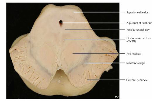
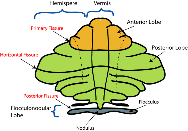
The cerebellum is a solid large structure attached to each side of the pons and separated from the dorsal surface of the pons by the fourth ventricle.

The cerebellar cortex is deeply grooved and appears as thin, transverse leaves called folia which run horizontally from one side to the other. While left and right hemispheres are recognised, they form a continuous solid structure, joined in the mid-line by the vermis..

The cerebellum has several lobes. The posterior lobe developed greatly when humans evolved and adopted the upright posture; at the same time the forebrain expanded and there are important connections between these two.

The older parts of the cerebellum include the flocculonodular lobe, which has connections with the vestibular apparatus and is particularly concerned with balance and eye movements.

The cerebellum is concerned with the control of movement, integrating signals from different parts of the nervous system and generating error signals that allow adjustments to be made to achieve the desired objective of the movement. In cerebellar disease, voluntary movements can become grossly exaggereated and attempts to correct them result in a tremor that occurs during voluntary movement.

Abnormal eye movements, nystagmus, can also appear in cerebellar disease.

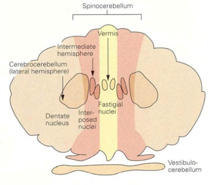
The cerebellum is divided into two hemispheres, one on either side of a central region called the vermis.

The cerebellar cortex is deeply grooved and appears as thin, transverse leaves called folia which run horizontally from one side to the other.

The left and right hemispheres form a continuous solid structure, joined in the mid-line by the vermis..

The cerebellum has can be divided into several lobes. The posterior lobe developed greatly when humans evolved and adopted the upright posture; at the same time the forebrain expanded and there are important connections between these two.

The older parts of the cerebellum include the flocculonodular lobe, which has connections with the vestibular apparatus and is particularly concerned with balance and eye movements.

The cerebellum arches over the pons from which it is separated by the fourth ventricle.

Connections between the brain and the cerebellum pass through the cerebellar peduncles, which are sometimes divided into 3 sections: the inferior, middle and superior cerebellar peduncles.

*


More on the Cerebellar Pathways Deep Cerebellar Nuclei
Cerebellar Peduncles
Overview of Cortico-Cerebellar Circuitry
The Red Nucleus and its projections
The Olive and its connections

"BodyParts3D, © The Database Center for Life Science licensed under CC Attribution-Share Alike 2.1 Japan." via Wikipedia Commons
Nrets at en.wikipedia
what-when-how.com

Top


Development of the Cerebellum

During the course of evolution, three areas of the cerebellum became involved with specific functions and these areas have retained the connections established at each stage of evolution.

The oldest part of the cerebellum is the flooculonodular lobe, situated caudally, behind the main mass of the cerebellar hemispheres; it is sometimes called the archicerebellum. This region is concerned with balance and eye movements, and receives important information from the vestibular apparatus that it uses to control eye movements in such a way as to preserve an image of an object on the fovea of the eye when the head moves in any direction. It is separated from the posterior lobe by the postero-lateral fissure, and its output pathways is through the fastigial nuclei.

A second older part of the cerebellum is the vermis and the anterior lobe which receives information about body position via the spino-cerebellar tracts, and communicates with the spinal cord and the muscles of the body to regulate muscle tone and posture. It is separated from the posterior lobe by the primary fissure, and is sometimes called the spinocerebellum (paleocerebellum); its output pathway is through the interposed nuclei (N interpositus; or Globose and Emboliform nuclei).

*

The third main area of the cerebellum, sometimes called the neocerebellum or cerebro-cerebellum, is large and developed in the lateral parts of each hemisphere during the period when primates attained the upright posture. At this stage there was great enlargement of the cerebral and cerebellar hemispheres and the development of essential connections between the cortex, pons and cerebellum. The output pathway is through the large Dentate nuclei. This part of the cerebellum, together the the motor areas of the cerebral cortex, is concerned with the planning and the dynamics involved during the execution of movements. Some areas of the parietal cortex concerned with visual guidance of movements, also project to the neocerebellum.

Quizlet.com

Top

Gross anatomy of the Basal Ganglia

Components of the Basal Ganglia

The main components of the basal ganglia are the striatum (caudate nucleus and putamen), the globus pallidus, the substantia nigra, the nucleus accumbens, and the subthalamic nucleus.

The striatum is the largest component of the basal ganglia and is the largest of the nuclei within the cerebral hemisphere. The term "striatum" comes from the observation that this structure has a striped appearance when sliced in certain directions, arising from numerous large and small bundles of nerve fibers (white matter) that traverse it.

The caudate nucleus is a part of the striatum with similar functions and can be considered as two separated parts of a single entity. They are separated anatomically by the internal capsule.

The striatum or putamen (also sometimes called the lentiform body or lenticular nucleus in older texts) receives input from many brain areas but sends output only to other components of the basal ganglia.

The globus pallidus is adjacent to the striatum and receives input from it. It is paler in sections than the putamen, hence its name.

The substantia nigra is a dark pigmented band within the cerebral peduncle and projects to the striatum.

The subthalamic nucleus, found in the diencephalon, receives input mainly from the striatum and cerebral cortex, and projects to the globus pallidus.

*


The corpus striatum can be seen to be a lens shaped body attached at its anterior pole to the head of the caudate nucleus. Posteriorly these two structures are separated by the internal capsule, as the caudale nucleus assumes a 'C' shape and swings into the temporal lobe, where it ends in the amygdaloid body.

The corpus striatum sits in a lateral position to the internal capsule, which in turn is lateral to the thalamus. The internal capsule passes between the corpus striatum and the caudate nucleus

Each of these areas has a complex internal anatomical and neurochemical organisation.

Substantia Nigra

Below and behind the thalamus and basal ganglia lies the midbrain and within the cerebral peduncles (crus cerebri), the ventral part of this structure on each side, can be seen a band on dark material known as the substantia nigra. This has important connections with the basal ganglia, and the cells that contain both the dark pigment and the neurotransmitter dopamine degenerate in Parkinson's disease


More on the Internal Structure of the Basal Ganglia

Internal Structure of the Corpus Striatum
The Substanita Nigra
The Subthalamic Nucleus


webspace.ship.edu


Chapter 5 : Cerebellum and Basal Ganglia

Brain: Contents Page

The Cerebellum and Basal Ganglia                         Topics :     

© HumanPhysiology.Academy 2014-2015